为什么生物的感知器官都集中在头部?
 为什么生物的感知器官一般都集中在头部?
 vx: wenmiau
这个问标题问题前没有一致的权势巨子诠释,这里我仅供给一些假设和证据。
拿网线[1]做一个类比,我们常用的网线有 cat3(三类),Cat5(五类), Cat5e(超五类), Cat6(六类), Cat6a(超六类)等几个尺度,按照挨次,其传输速度越来越高,同时其有用传输距离越来越短:
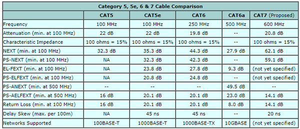
按照传输速度,千兆收集一般保举利用超五类及以上彀线。同时,跟着尺度的提高, 响应速度越来越快;为包管传输速度(噪声按捺),网线长度越来越短,单元长度的当作本也越来越高
高标网线做的短、粗、屏障好、线束多是由噪声按捺、响应速度、传输速度等几个方面的身分决议的。之所以能拿网线作为类比,是因为作为感知信息传递的载体,神经纤维束也存在着这些机能要求:传输带宽、响应速度、噪声按捺[2]. 神经纤维 / 神经纤维束素质上传递的是数 / 模夹杂旌旗灯号[3].
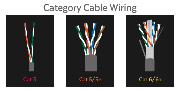
对于一个典型的由树突(Dendrites)、胞体(Soma)和轴突(Axon)构成神经细胞,其旌旗灯号传递的最长一段显然是轴突。因为神经旌旗灯号的生化反映素质,我们可以简单认为统一种神经细胞的单根轴突传递旌旗灯号的带宽是一致的(接近的)。
神经细胞根基布局:树突(Dendrites)、胞体(Soma)和轴突(Axon)
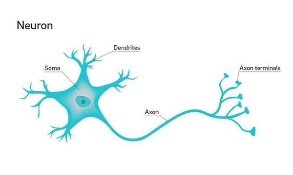
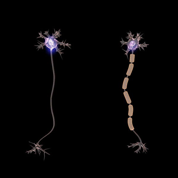
跟网线一样,有些轴突会有屏障层——髓鞘(Myelin Sheath)[4], 这对旌旗灯号传递过程中的噪声按捺具有积极感化:
轴突的屏障层——髓鞘(myelin sheath)
从神经旌旗灯号的整个传输和处置过程来看,在各个阶段噪声都有可能发生[5]:
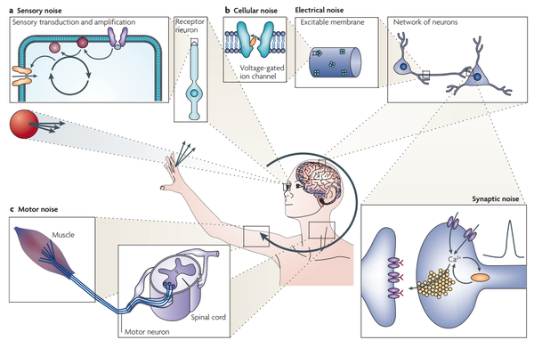
从感知到传输,再到旌旗灯号的处置(此中也需要按照处置阶段进行处置区域之间的传输),神经旌旗灯号城市混入噪声,这是由神经旌旗灯号的生化反映素质决议的[5].
轴突的屏障层——髓鞘(myelin sheath)
具体地,我们考查一段轴突上旌旗灯号在传递过程中的环境,可以不雅察到肉眼可见的跟着传输距离增添而噪声增大的环境[6, 7]:
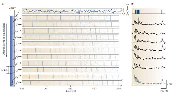
在轴突上每隔 150 微米进行旌旗灯号采样(a), 可以看到旌旗灯号传输过程中的转变环境(b)
不难理解地,噪声旌旗灯号,出格是传输过程中引入的噪声旌旗灯号对感知信息的处置具有负面的结果[8, 9]. 按照上面的信息,我们可以猜想,跟着感知旌旗灯号带宽和响应速度要求的增添,神经纤维束(树突的调集体)越粗、而且越短,其髓鞘屏障越好。
以旌旗灯号带宽最高的感知旌旗灯号——视觉旌旗灯号为例()[10]:
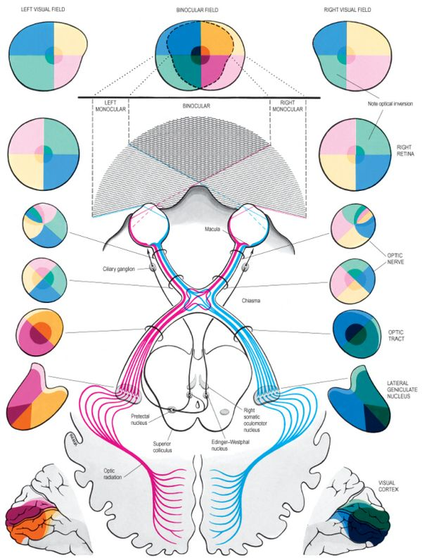
视觉旌旗灯号从眼睛底部起头就进入大脑,然后经由过程视神经(optic nerve)等布局进行传输和处置(边传输边处置)[10]
严酷意义上,视觉旌旗灯号被视网膜感知就已经进入了“大脑”,然后经由过程视神经(optic nerve)进行传输。是以视神经中传输的是最为原始的,根基未经大脑进行感知加工过的旌旗灯号。其直径是单一功能神经纤维中最粗的[11, 12].
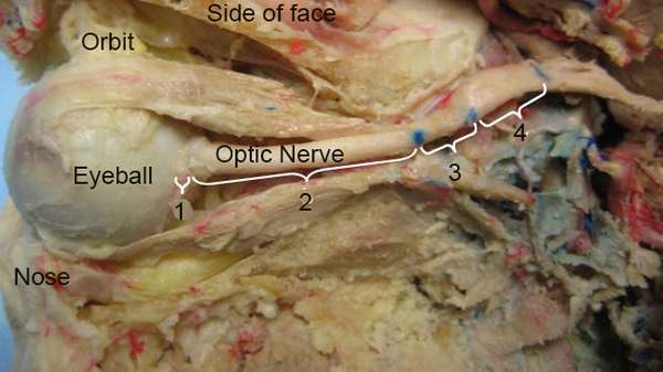
视神经剖解环境,可见其粗细
同时,视神经具有较粗的髓鞘作为屏障层庇护旌旗灯号的传输[13]:
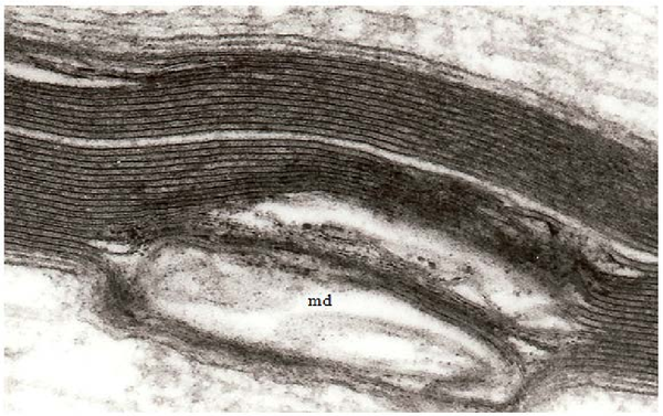
正常的视神经髓鞘(上部)和受损的髓鞘(下部, md)
具体地,对于人类来说,视神经髓鞘的损坏将直接影响人的视觉感知,好比视神经炎、多发性硬化等城市损坏人的视觉[14].
若是以数字旌旗灯号来等效,视神经的一般传输速度为约 8.75Mb/s, 而且具有峰值约 40Mb/s 的传输能力[15], 这根基上是 Cat5 网线的尺度。巧合的是,二者的粗细也是差不多的。比拟之下,传输嗅觉旌旗灯号的嗅神经纤维就要细得多了[16], 因为其带宽要求也低得多。
在传输速度(响应速度)上,神经旌旗灯号约为 100m/s[17]. 不外具体数值受到良多身分的影响,好比春秋、性别、疾病,以及神经纤维的粗细和髓鞘[18, 19]:
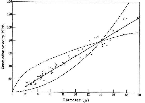
神经纤维越粗,神经旌旗灯号传递越快
髓鞘环境(旌旗灯号屏障)越好,神经旌旗灯号传递越快
是以,像视觉、听觉、嗅觉、味觉这样对传输带宽(旌旗灯号丰硕性)、响应速度或者噪声按捺(敏感性)要求高的旌旗灯号,从旌旗灯号发生到旌旗灯号处置之间的距离短,对生物的保存和演化具有更为积极的意义。这些感知器官离大脑近的生物种类和个别,是以在天然选择中具有竞争优势。
[1] McNeill, D. B., & Kilpatrick, J. B. (1995).U.S. Patent No. 5,399,813. Washington, DC: U.S. Patent and Trademark Office.
[2] Furness, J. B. (2006).The enteric nervous system(Vol. 274). Oxford: Blackwell.
[3] Clark, B., & Häusser, M. (2006). Neural coding: hybrid analog and digital signalling in axons.Current biology,16(15), R585-R588.
[4] Michailov, G. V., Sereda, M. W., Brinkmann, B. G., Fischer, T. M., Haug, B., Birchmeier, C., ... & Nave, K. A. (2004). Axonal neuregulin-1 regulates myelin sheath thickness.Science,304(5671), 700-703.
[5] Faisal, A. A., Selen, L. P., & Wolpert, D. M. (2008). Noise in the nervous system.Nature reviews neuroscience,9(4), 292.
[6] Fellous, J. M., Tiesinga, P. H., Thomas, P. J., & Sejnowski, T. J. (2004). Discovering spike patterns in neuronal responses.Journal of Neuroscience,24(12), 2989-3001.
[7] Steriade, M., McCormick, D. A., & Sejnowski, T. J. (1993). Thalamocortical oscillations in the sleeping and aroused brain.Science,262(5134), 679-685.
[8] Neishabouri, A., & Faisal, A. A. (2014). Axonal noise as a source of synaptic variability.PLoS computational biology,10(5), e1003615.
[9] Maham, B. (2015). A communication theoretic analysis of synaptic channels under axonal noise.IEEE Communications Letters,19(11), 1901-1904.
[10] Gray, H., & Standring, S. (2008).Gray's anatomy: the anatomical basis of clinical practice. Churchill Livingstone.
[11] Mikelberg, F. S., Drance, S. M., Schulzer, M., Yidegiligne, H. M., & Weis, M. M. (1989). The normal human optic nerve: axon count and axon diameter distribution.Ophthalmology,96(9), 1325-1328.
[12] Vaiman, M., Abuita, R., & Bekerman, I. (2015). Optic nerve sheath diameters in healthy adults measured by computer tomography.International journal of ophthalmology,8(6), 1240.
[13] Maxwell, W. (2013). Damage to myelin and oligodendrocytes: a role in chronic outcomes following traumatic brain injury?.Brain sciences,3(3), 1374-1394.
[14] Klistorner, A., Arvind, H., Nguyen, T., Garrick, R., Paine, M., Graham, S., ... & Yiannikas, C. (2008). Axonal loss and myelin in early ON loss in postacute optic neuritis.Annals of Neurology: Official Journal of the American Neurological Association and the Child Neurology Society,64(3), 325-331.
[15] Koch, K., McLean, J., Segev, R., Freed, M. A., Berry II, M. J., Balasubramanian, V., & Sterling, P. (2006). How much the eye tells the brain.Current Biology,16(14), 1428-1434.
[16] Gasser, H. S. (1956). Olfactory nerve fibers.The Journal of general physiology,39(4), 473-496.
[17] Hursh, J. B. (1939). Conduction velocity and diameter of nerve fibers.American Journal of Physiology-Legacy Content,127(1), 131-139.
[18] Smith, R. S., & Koles, Z. J. (1970). Myelinated nerve fibers: computed effect of myelin thickness on conduction velocity.American Journal of Physiology-Legacy Content,219(5), 1256-1258.
[19] Waxman, S. G., & Bennett, M. V. (1972). Relative conduction velocities of small myelinated and non-myelinated fibres in the central nervous system.Nature New Biology,238(85), 217.
- 发表于 2019-04-16 23:23
- 阅读 ( 1443 )
- 分类:其他类型
0 篇文章
作家榜 »
-
 xiaonan123
189 文章
xiaonan123
189 文章
-
 汤依妹儿
97 文章
汤依妹儿
97 文章
-
 luogf229
46 文章
luogf229
46 文章
-
 jy02406749
45 文章
jy02406749
45 文章
-
 小凡
34 文章
小凡
34 文章
-
 Daisy萌
32 文章
Daisy萌
32 文章
-
 我的QQ3117863681
24 文章
我的QQ3117863681
24 文章
-
 华志健
23 文章
华志健
23 文章